
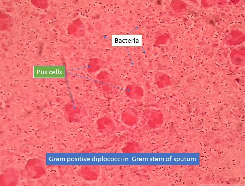
Gram Negative Rods 3. 1 A Gram stain from a carefully collected specimen with neutrophils and lancet-shaped diplococci staining gram-positive which are intracellular or encapsulated can provide strong support to the clinical diagnosis of pneumococcal pneumonia.
Gram Positive Diplococci Introduction Pathogenecity Lab Diagnosis

Gram Stain Of Blood Cultures Demonstrating Gram Negative Diplococci In Download Scientific Diagram
Streptococcus Pneumoniae
Gram in 1884 it remains an important and useful technique to this day.
Gram positive diplococci. The name originates from the word diplo meaning double and coccus meaning berry. Gram Negative Coccobacilli. Gram stain and bacterial morphology.
These small pleomorphic gram-negative bacteria range in shape from round cocci to short thin rods bacilli. Cell Shape and Arrangements. Non-spore forming Some species non-human isolates are positive.
Streptococcus pneumoniae infects the human anatomy in the respiratory tract and the immune system. For many years the retention of Gram stain was one of. In contrast to simple stains differential stains are used to distinguish the difference between bacteria.
The two coccus cells are connected. Growth of a gram-negative oxidase-positive diplococcus from the urethra men or endocervix women on a selective culture medium and demonstration of typical colonial morphology positive oxidase reaction and typical gram- negative. Biochemical Test and Identification of Neisseria gonorrhoeae.
Streptococcus bacteria is Gram-positive and are generally spherical in shape. Streptococcus pneumoniae is a gram-positive encapsulated lancet-shaped diplococci most commonly causing otitis media pneumonia sinusitis and meningitis. One of the most well-known differential stains is Gram stain which differentiates gram-positive and gram-negative bacteria based on the difference in their cell wall structure.
The Gram stain is a differential staining technique used to classify categorize bacteria into two major groups. The Gram stain was developed by the Danish bacteriologist Hans Christian Gram. Its size ranges between 0608 μm.
Gram Positive Cocci 2. Presence of Gram-negative diplococci in phagocytes of the central nervous system Treatment Penicillin administered intravenously is the drug of choice Prevention Eradication is unlikely due to the presence of asymptomatic carriers High risk vaccine Diagnosis Treatment and Prevention. One example of diplococci strains is Neisseria gonorrhea.
Some streptococci such as Streptococcus pneumoniae Pneumococcal Infections Streptococcus pneumoniae pneumococci are gram-positive alpha-hemolytic aerobic encapsulated diplococci. Are a Gram-negative non-spore-forming diplococcus that has a flattened shape. Streptococcus Bacteria Classification Shape Infection Gram Stain Overview.
Gram positive rods Gram negative rods Gram postive cocci and Gram negative cocci see images below. Gram positive cells are simpler chemical structure with a acidic protoplasm. They can reach a diameter of around 0608 μm and sometimes up to 10 2030 μm.
Crystal violet the primary stain of the Gram stain procedure is readily retained and stabilized within this matrix causing gram-positive prokaryotes to appear purple under a brightfield microscope after Gram staining. They are gram -ve non-haemolytic catalase and oxidase both positive organism. In the US pneumococcal infection is a major cause of otitis media pneumonia sepsis.
Morphology cocci bacilli coccobacilli spiral or presence of branching filaments Gram-staining properties Grampositive Gramnegative and atypical metabolic activity aerobic anaerobic microaerophile or facultative or virulence factors eg presence of capsule pili proteins formation of. A typically gram-negative bacterium causes a sexually transmitted infection called gonorrhea. Prokaryotes are identified as gram-positive if they have a multiple layer matrix of peptidoglycan forming the cell wall.
Diplococcus bacteria diplococci are arranged in pairs ie. Results obtained by culture without evaluation for contamination may be noncontributory or misleading. A differential stain like that invented by Hans Christian Gram in 1882 will give you more information and allow you to group the stainable bacteria into more groupings.
Rods or Bacillus 1. A coccobacillus is a type of bacterium with a shape intermediate between cocci and bacilli ie they are very short rods that may be mistaken for cocci. Mutans and Strep mitis found in the normal flora of the oropharynx commonly cause dental carries and subacute bacterial endocarditis Strep.
They can cause gonorrhea Neisseria gonorrhoeae pneumonia Diplococcus pneumoniae. Crystal violet the primary stain of the Gram stain procedure is readily retained and stabilized within this matrix causing gram-positive prokaryotes to appear purple under a brightfield microscope after Gram staining. Human pathogenic bacteria can be classified according to their characteristics.
Prokaryotes are identified as gram-positive if they have a multiple layer matrix of peptidoglycan forming the cell wall. They are oxidase-positive non-acid-fast cocci or plump rods. This bacteria congregates at the end of the bronchial tubes in the alveoli which elicits an inflammatory results.
Single Cell Coccus Spiral Curved Palisades Chains Pair of Cells Diplobacilli Coccobacillus Tetrads Clusters Chains Pair of Cells Diplococci Single Cell Bacillus DIVISION OF LABORATORY SYSTEMS. Hence the bacteria are called coccobacilli. The Gram staining is one of the most crucial staining techniques in microbiology.
Of all the different classification systems the Gram stain has withstood the test of time. Diplococci chains streptococci groups of four tetrads or eight sarcina or grape like clusters staphylococci. However culture of expectorated sputum in.
It allows a large proportion of clinically important bacteria to be classified as either Gram positive or negative based on their. Cocci in grape-like clusters. Examples of gram-positive diplococci pathogens include Streptococcus pneumoniae and some species in Enterococcus bacteria.
Differential stains- Gram stain. Gram reaction of the bacteria whether Gram-positive or Gram-negative Morphology of the bacteria whether cocci diplococci streptococci rods or. This type of bacteria may be gram-positive or gram-negative.
These bacteria are usually found in pairs as two joined cells. Hanna in Encyclopedia of Food Microbiology 1999 Bacterial Characteristics. It gets its name from the Danish bacteriologist Hans Christian Gram who first introduced it in 1882 mainly to identify organisms causing pneumonia1 Often the first test performed gram staining involves the use of crystal violet or methylene blue as the primary color2 The term for organisms that retain the.
For many years the retention of Gram stain was one of. Neisseria gonorrhoeae also known as gonococcus singular or gonococci plural is a species of Gram-negative diplococci bacteria isolated by Albert Neisser in 1879. It has a thick peptidoglycan layer.
Gram Variable Rods. Gram positive and Gram negative based on the differences of the chemical and physical properties of the cell wall. They are commonly found in the mucous membrane of the mouth and respiratory tract etc where they have been associated with a number of diseases and infections including sepsis pneumonia and pharyngitis.
It causes the sexually transmitted genitourinary infection gonorrhea as well as other forms of gonococcal disease including disseminated gonococcemia septic arthritis and gonococcal ophthalmia neonatorum. Typical gram-negative intracellular diplococci on microscopic examination of a smear of urethral exudate from men or endocervical secretions from women. Streptococcus viridans consist of Strep.
Teichoic acids are intertwined among the.
Gram Staining Rules

Pneumococcus Introduction Morphology Pathogenecity Laboratory
Gram Negative Diplococci Images Stock Photos Vectors Shutterstock

Gram Positive Organisms Flashcards Quizlet
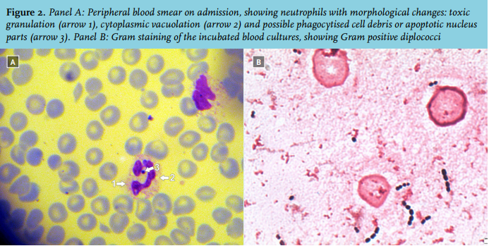
Article Answer To Photo Quiz A Blood Smear On Admission Full Text November 2017 Njm

Cns Pathology
Wiv Isp Be

Gram Stain Showing Gram Negative Diplococci In Thrombus Download Scientific Diagram


